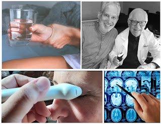
COGNITION TESTING & CONCUSSION METRICS THROUGH OPTICSCAN
INTRODUCTION A new research study is underway at the Bard Diagnostic Imaging Center in NYC. Dr. Robert Bard and a local research team combines the latest biometric scanning advantages of transcranial DOPPLER imaging, hemodynamic ULTRASOUND scanning (of the retinal, basilary and temporal arteries) and the integration of BIOFEEDBACK technology for a special research study. This neuro-scan review establishes (1) a cross-comparative study between the technologies and (2) a multi-modality screening & monitoring protocol to record post-concussion and neurodegenerative disorders.
One of several major foci for this project is to explore the development of a diagnostic paradigm in the detection of post concussion symptoms including the growing reports of Chronic Traumatic Encephalopathy/CTE, identified from head concussions in military service and high impact sports. Since 2018, Dr. Bard has been collaborating with neurologists and pain management specialists to assess the rampant growth of this progressive brain condition.
STUDY 1: INTRACRANIAL PRESSURE
Case studies with a presumptive degenerative neuromuscular disease or amyotrophic lateral sclerosis are now being examined through a non-invasive investigation of the eye. We can observe increased intracranial pressure, which may reflect in changes in the optic nerve diameter.
[Fig. 1] Scan of the eye (L) with a Doppler ultrasound probe shows the optic nerve diameter of five millimeters compared to the usually symmetric right almost eight millimeters. This suggests increased intracranial pressure or optic nerve tumor indicating a blood flow measure of the right anterior cerebral artery circulation that supplies the retina (approximately 60cm/s) blood flow. [Fig. 2] The left vessel shows a decreased pressure of approximately 45cm/s as shown by the decreased height of the blood flow graph at the bottom. These quantitative diagnostic technologies are non-invasive allowing close clinical follow-up treatment in diseases affecting the eye related to brain trauma or degenerative neuromuscular disorders.
STUDY 2: POST-CONCUSSIVE / RETINAL ARTERIOVENOUS IMAGING
Concussion literature notes a funduscopic exam of the retinal arterial-venous width showing ASYMMETRY is indicative of possible trauma. This is a potential response of the autonomic nervous systems microcirculatory hemodynamics that are optically useful in validating the treatment progress. [L Image] Normal retinal artery vein width ratio is 1 to 2. [R image] artery 0.9 mm (red) vein 2.3mm (blue) signifying possible increased intracranial pressure. Hemodynamic Doppler study of abnormal retinal vessels allows real time evaluation of the cerebral circulation measuring the anterior cerebral artery (ACA) with physiologic flow parameters.
STUDY 3: WHIPLASH
Hemodynamic changes from whiplash may injure the vessels supplying the brain. [Image L] High resolution scan of traumatized carotid artery showing turbulent flow (dark blue) and normal outer wall (red) ruling out arterial wall rupture. [Image R] blood flow in the vertebral artery shows less flow disturbance (scattered blue) and no evidence of thrombosis which is a common cause of posttraumatic symptoms including speech and gait abnormalities
Now that more radiologists are using Doppler blood flow to examine eye disease, including systemic diseases (including diabetes, brain tumors, heart disease, sickle cell disease, etc.) that affect the eye, we are hopeful that the ophthalmologic and neurological communities will start using this noninvasive technology as well to improve noninvasive and more rapid treatment of potential eye disorders, such as cancers of the eye, diabetes and glaucoma. Another future use will be to correlate the effect of decreased vascular pulsation in the production of cerebrospinal fluid that is removed by the cleansing glymphatic system is postulated as a contributing factor in degenerative neuromuscular disease.
BLOOD FLOW STUDY 101: Hemodynamics is defined as the study of blood flow in relation to the status of the circulatory system and homeostatic mechanisms of autoregulation. Through the monitoring of blood flow, diagnostic analysis provides answers to the health and physiological status of the target area scanned as well as cell-level metabolism, regulation of pH, osmotic pressure and protection from microbial and mechanical harm. Assessing injuries, inflammation or mutative growths by assessment of blood flow allows diagnostic criteria regarding the severity of tissue disorders or tumor malignancy.
3D DOPPLER ULTRASOUND: A MAJOR ASSET TO RESEARCH
From simple case studies to double blind clinical trials, the many benefits of non-invasive imaging offers repeatable, visual confirmation of treatment efficacy. Ultrasound is the most widely used diagnostic technology worldwide and, in particular, 3D Doppler histogram analysis is targeted to collect a patient's biometric data safely and efficiently, thanks to its vastly improved quantitative reporting capacity.
Under exploratory device tech reviews, this video shows the effects of electromagnetic pulse wave neuro-stimulation and the induction of improved microcirculation on the body that are some noninvasive modalities that are easily monitored with an ultrasound scan using Doppler or elastography modalities. In the case of electromagnetic devices, the involuntary muscle contraction is evidence of the electrical changes in the targeted muscle developed by this technology which continues to find new evidence, supporting its ability to recover the body's healing process through cellular regeneration on a preliminary study, quantitative measurement that the regenerative timeline through the use of a neurostimulator through a simple before and after comparison shows the body's reaction to the therapeutic device.
THE GUT-BRAIN CONNECTION
By: Dr. Robert L. Bard / Dr. Leslie Valle
The intestinal system is now recognized as a major factor in the immune system. New ultrasound imaging advances allow imaging of the gut wall layers, internal vasculature and areas of inflammation and post inflammatory fibrosis with stricture or deformation. High resolution probes visualize the various segments of digestive organs for scar formation and strain elastography or more recently shear wave elastography quantifies the degree of fibrosis. This exam may be performed endoscopically or by transabdominal probes depending on the location of the organ and type of pathologic process. Certain areas, such as the appendix, are quickly evaluated for appendicitis with high accuracy on CT studies.
Other organs like the pancreas are examined with endoscopic MRI for silent cancers and regional adenopathy. Inflammatory markers of active disease are studied with Doppler flow hemodynamics including 3D measurements of the inflammatory arteries attacking the swollen bowel wall. Disease such as Crohn’s disease, irritable bowel syndrome (IBS) and diverticulitis are accompanied by an influx of feeding vessels. Greater arterial flow in the affected area signifies the likelihood of increased bowel involvement leading to exterior wall adhesions possibly obstructing or strangulating adjacent healthy small bowel loops. Fistulae, abscess and cancers may be observed with real time sonographic evaluation in quick non-invasive surface scanning sessions or with surgical intraoperative imaging for locoregional metastases.
ELASTOGRAPHY: ADVANCING ULTRASOUND TO SCAN THE GUT
Developed in Europe, Great Britain and Japan 20 years ago sonography of the GI tract was slow to be adopted in the US. As of 2 years ago, only one center in New York City was performing outpatient elastography for colitis, IBS and Crohn’s disorder. There are many levels of sophistication in Doppler flow imaging as well as elasticity measurements and the clinical subtype will guide the probe application and recording options. Wide usage in cancer detection of the thyroid, liver, pancreas, breast, prostate and skin has made screening popular worldwide. Often a biopsy will be avoided by a negative elastogram report in many developed nations. Cancers are hard and inflammation is soft which are readily observable endpoints on most elastographic studies. Elastography is applicable to examining the pediatric brain as sound easily penetrates the younger skull tissue and is actively used in diagnosing pediatric medulloblastoma and neuroblastoma.
ON POST-CONCUSSION SYMPTOMS
The underreported dermal accumulation and bowel permeability to toxins and the physiologic gastroinstestinal effects of concussion is likewise mostly anecdotal. The possibility of scanning the gut for increased blood flow following brain trauma may be easily performed and sequentially followed as a guideline for evaluation of concussion’s traumatic chronicity. As the bowel activity is regulated by the autonomic nervous system we may apply the same diagnostic endpoints as other physiologic norms. The response of the microvasculature in the retina provides a functional guideline as to the progression of brain trauma and concussion as measured by blood flow. The application of optical devices and sensors for physical (temperature, respiration, heart rate, blood pressure) chemical (pH, pO2, glucose, lipids, oximetry) and biological (antigens, antibodies, electrolytes, enzymes, inhibitors, metabolites, proteins) data with imaging (endoscopy, optical tomography, confocal microscopy, laser speckle spectrometry) adds potential new classifications of metabolism in the pathologically altered state. The addition of wearable sensors and portable laser powered LED tools currently used by athletes are applicable to non invasive monitoring of the effect on the body by concussion indirectly. Surrogate markers are widely used such as analyzing the white blood cells to verify the infection is bacterial rather than viral. Similarly, there exists alternate options with high accuracy to move forward the immediate and long term effects of concussive injury.
Sunday, June 19, 2022 - One of the most exciting opportunities in neuroscience research today is the use of strategies that protect the brain which may potentially prevent, delay or inhibit the progression of neurodegenerative diseases such as Alzheimer’s, Parkinson’s and ALS. This opportunity rests on our ability for early diagnosis. Research has shown that the likelihood of success for a given treatment-whether lifestyle changes or pharmacological approaches- is highly dependent upon early intervention, before the disease process has become too severe and potentially irreversible. Therefore, it is critical to understand the underlying biological changes that occur in the early stages of these diseases, along with the technology to detect them. (See complete article)
VALIDATION WITH DIAGNOSTIC IMAGING
Since inception, the acceptance of non-invasive imaging for surgical guidance, monitoring pathology or screening has been a game-changer in just about every area of medicine. Through the AngioFoundation (501c3) and other non-profit research partnerships, Dr. Robert L. Bard (cancer radiologist and diagnostic imaging specialist) has helped deploy countless exploration studies, case studies and clinical trials in support of non-invasive diagnostic solutions while validating (or challenging) therapeutic claims. As a researcher, educator and technology advocate, Dr. Bard is also co-founder and associate publisher of a non-commercial program called "MedTech Reviews"- a public newsletter where he shares his clinical expertise in diagnostic interpretation / analyses in performance reviews of health technologies . All results are based on a BEFORE-AND-AFTER visual comparison and a clinical interpretive description of the imaging graphic - thus aiming to show the body's potential reaction to that device (if any) by virtue of an after-treatment applied scan. (see: BardClinicalTrials.com)
Traumatic brain injuries can contribute to both short-term and long-term issues with cognitive function, but they can also impact emotional and physical health beyond the brain itself. While much of the research to date has focused on more severe forms of traumatic brain injury, it is now expanding to evaluate concussions.
Writing this article reminded me that I, too, am part of this story. I had two episodes of concussion in my teens and early twenties, neither related to sports: one from a fall where I hit my head, another from a car accident that resulted in whiplash. In both of those cases, I was just told to rest until my head stopped hurting and then resume normal activity. Fortunately, I recovered without any long-term issues. In the decades since then, our understanding of head injuries has greatly expanded, prompting innovations in both diagnosis and treatment.
Concussions are viewed as a mild form of traumatic brain injuries and most frequently occur following an event that involves an acceleration–deceleration mechanism without actual injury to the head, such as whiplash, or the head striking an object. As we study these, researchers and clinicians are learning that these are fairly common, but often underdiagnosed.
According to the CDC, an estimated 1.6 – 3.8 million people suffer from concussions related to sports or recreational activities every year. A National Health Interview Survey in 2020 found that 6.8% of children aged 17 years and under had ever had symptoms of concussion, while only 3.9% had ever been diagnosed. [1] There is also good evidence to suggest that an athlete who has had one concussion is also more likely to suffer from multiple concussions and suffer long-term consequences. [2] Not all head injuries occur in athletes, but these are the most studied.
While the vast majority of people with concussions recover without obvious disability, people can end up with long-term cognitive, emotional and functional issues affecting quality of life – including memory issues and Alzheimer’s disease. [3] Efforts to better predict outcome from head injuries by focusing on the age, sex, type of injury and acute assessments have led to some improvement, but still fail to predict or explain the variation in healing and outcomes.
Studies in professional athletes have shown that about 80–90% are sufficiently recovered to return to playing within 7–10 days. But that means that 10-20% are not, and their recovery can take up to 3 times longer. Even taking into account variations in initial injury, this variation is difficult to explain or predict. [4]
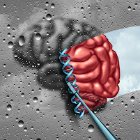
DNA RESEARCH LINKS TO INJURY & HEALING RESPONSE
Brain injury is broken down into two phases: a primary phase and a secondary phase. The primary phase is the result of the physical or mechanical forces on the brain causing direct injury. The secondary phase involves the brain’s response to the injury – a complex interplay of multiple biological systems including immune, vascular, neuroendocrine, neurotransmitters, neuroplasticity and even mitochondria and epigenetics. [3] In concussions, it is typically this secondary phase that plays a major role in how well an individual responds and recovers – both in time and function.
DNA is the genetic code that is the blueprint for everything that goes on in our bodies. Genomics is the study of how small changes in our DNA affect how our bodies function. [See feat. on Genomics testing] Research, primarily focused on combat veterans and athletes so far, has shown that these small variations in our DNA may account for at least some of why some people respond to and recover from traumatic brain injury better than others.
The APOE gene plays many roles, including immune response and neuroplasticity. Carriers of the APOE4 gene can be predisposed to worse outcomes after traumatic brain injuries, especially if they are moderate or severe, or there are multiple concussions. While the APOE gene is the most widely studied, there are now over a dozen others that have been identified. Variants in other genes involved in the inflammatory response, blood flow, DNA repair, neuroplasticity, learning and memory are also implicated, including TNF alpha, IL1, IL6, NOS3, ACE, COMT, NMDA receptors, BDNF, KIBRA, MAPT, PARP, MME, SLC17A7, GRIN2A. Because there are hundreds of genes impacting all of these biological systems, it is likely that there are many to be still evaluated, and outcomes are the result of the interaction of multiple genes.
As genomics contributes to our understanding of how and why individuals can vary greatly in their ability to recover from traumatic brain injuries, it is paving the way for more personalized prevention and treatment strategies for concussions. Having accessible and noninvasive technologies to provide evaluation of brain injury and ongoing recovery will be a key part of this progress.
References: (1) https://www.cdc.gov/traumaticbraininjury/concussion/index.html (2) McCrory et al. Consensus statement on concussion in sport—the 5th international conference on concussion in sport held in Berlin, October 2016. Br J Sports Med 2017;0:1–10. (3) Bennett et al. Chapter 9: Genetic Influences in Traumatic Brain Injury, in Laskowitz D, Grant G editors. Translational Research in Traumatic Brain Injury. CRC Press/Taylor and Francis Group 2016. (4) Jane McDevitt & Evgeny Krynetskiy. Genetic findings in sport-related concussions: potential for individualized medicine? Concussion 2017; 2(1).
ROBERTA KLINE, MD (Educational Dir. /Women's Diagnostic Group) is a board-certified ObGyn physician, Integrative Personalized Medicine expert, consultant, author, and educator whose mission is to change how we approach health and deliver healthcare. She helped to create the Integrative & Functional Medicine program for a family practice residency, has consulted with Sodexo to implement the first personalized nutrition menu for healthcare facilities, and serves as Education Director for several organizations including the Women’s Diagnostic Health Network, Mommies on a Mission. Learn more at https://robertaklinemd.com/
July 19, 2022- IPHA NEWS conducted a private interview with the co-developers of NEUROVINE - a portable headband using EEG (electroencephalogram) technology to measure brain waves. Meet CEO Ashleigh Kennedy, Ph.D., and CMO Matthew Kennedy, MD, MSc (co-founders of Neurovine) from Ottawa, Ontario, CA. who shares their objectives in support of concussion monitoring by measuring brain health as part of optimizing their recovery process. Neurovine offers this portable interactive monitoring program for athletes, students, professionals and anyone undergoing mentally strenuous work by “alerting them to take brain breaks before an activity becomes too strenuous”.
Pain is everywhere. Billions of dollars are spent each year in the United States alone on pain treatments and remedies. Per the National Center for Health Statistics almost 60% are living with pain. While most sufferers of pain want relief and answers, many medical professionals may be providing inefficient or incorrect pain reliefs counters. The “cookie cutter” approach for pain over the last several decades has left the world in a pain crisis. One of the main reasons why- the current treatment model does not do an adequate job in categorizing pain. Pain falls under different categories. Most people do not understand that the type of pain you have directly correlates the type of treatment given. Personalized medicine in the treatment of pain holds the key to understanding the types of pain and developing the correct course of action to treat the pain.
A PARTNERSHIP WITH SPORTS MOMS
Another key launch pad to this research program is his alliance with MOMS OF ATHLETES, co-architected by Dr. Roberta Kline(Women's Diagnostic Network) and Dr. Donna Febres. This educational advocacy group supports clinical care and preventive science to college level athletes. This unique alliance of professionals and moms are dedicated to finding the safest options in injury and pain care - with an emphasis on new solutions for concussion research and prevention. This includes the modern non-invasive (non-surgical) alternatives where possible. NCMOA also teams up with the professional pain and medical associations to collaborate on latest protocols and information on ground-breaking neurostimulation, electromagnetic, holistic/full-body solutions and wearable therapeutics plus the latest in non-radiation clinical imaging validation. (see complete details)
Disclaimer: MEDTECH REVIEWS* is a non-commercial user review of health-related technologies and is not intended as a marketing program for any device(s) featured in this video for evaluation. This presentation is for informational use only does not offer any direct medical claims whatsoever. Statements from all speakers herein are expressing their own unscripted views that do not reflect those of our producers. Always seek the advice of your physician or other qualified health care provider with any questions you may have regarding a medical condition or treatment.
Copyright Notice: The materials provided on this website/web-based article are copyrighted and the intellectual property of the publishers/producers (The NY Cancer Resource Alliance/IntermediaWorx inc. and The AngioFoundation). It is provided publicly strictly for informational purposes within non-commercial use and not for purposes of resale, distribution, public display or performance. Unless otherwise indicated on this web based page, sharing, re-posting, re-publishing of this work is strictly prohibited without due permission from the publishers. Also, certain content may be licensed from third-parties. The licenses for some of this Content may contain additional terms. When such Content licenses contain additional terms, we will make these terms available to you on those pages (which his incorporated herein by reference).The publishers/producers of this site and its contents such as videos, graphics, text, and other materials published are not intended to be a substitute for professional medical advice, diagnosis, or treatment. For any questions you may have regarding a medical condition, please always seek the advice of your physician or a qualified health provider. Do not postpone or disregard any professional medical advice over something you may have seen or read on this website. If you think you may have a medical emergency, call your doctor or 9-1-1 immediately. This website does not support, endorse or recommend any specific products, tests, physicians, procedures, treatment opinions or other information that may be mentioned on this site. Referencing any content or information seen or published in this website or shared by other visitors of this website is solely at your own risk. The publishers/producers of this Internet web site reserves the right, at its sole discretion, to modify, disable access to, or discontinue, temporarily or permanently, all or any part of this Internet web site or any information contained thereon without liability or notice to you.


.jpg)


