
Disclaimer: "HealthTech Reporter" / "MEDTECH REVIEWS" is a publishing program presenting a non-commercial, non-official user review of portable health-related technologies on the market. Our reviews are not intended as a marketing program for any device(s) featured in this article or video for evaluation. It is not a replacement for an official medical device verification and validation process for regulatory standards. This presentation is for informational use only and does not offer any direct medical claims whatsoever. Statements from all contributing speakers herein are expressing their own personal unscripted views as ANECDOTAL and they do not reflect those of our producers/publishers. Always seek the advice of your physician or other qualified health care provider with any questions you may have regarding a medical condition or treatment.
EVALUATION OVERVIEW: MULTI-MODALITY DEVICE TEST DRIVE / USER REACTIONS & IMAGING SCANS
Level 1 & 2 Case Study: Physiological Effects & Ultrasound Imaging

a. Evaluation on meditation effects on the body
b. Assessment and comments on the device's components (as stated in its user literature)
- binaural beats, isochronic tones, holographic music
- red and blue light / frequency therapy
c. Evaluation of the theories behind the device's design. Discussion on the variety of ways of boosting brain levels of serotonin, beta-endorphins and norepinephrine (as per the mentions of this device in social media, from other users blogs, literature and videos)
2) BEFORE & AFTER STUDY: To evaluate the effects of said device, Dr. Robert Bard co-designed the study strategy behind this Phase 1 review with evaluators including Jessica Glynn, LCSW. The review is an academic exercise in workflow analysis, comprised of a 9-point review from an unofficial personal study (not to be confused with a clinical trial or medical review), this report explains the possible means of gathering data averages from a limited number of subjects using medical-grade diagnostic modalities. The logic behind this review dictates collecting a BEFORE AND AFTER of scans and vitals for comparison, possibly linking the user's reaction(s) to the device.
* BEFORE (BASELINE) studies include:
a. Preliminary exam of the subject's VITAL SIGNS: Recording includes: BLOOD PRESSURE, BODY TEMPERATURE, HEART RATE AND BLOOD/OXYGEN SATURATION. These are identified to show reactive effects when they body is placed in a meditative state.
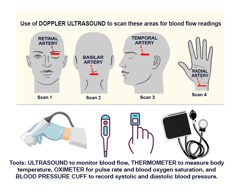
b. Ultrasound scan of the BASILAR ARTERY located in the back of the head/brain. This blood vessel supplies oxygen-rich blood to areas of the brain and the autonomic/central nervous system.
c. Ultrasound scan of the EYE/RETINA, specifically to study the optic nerve to show the paucity of the vessels in the central retinal artery and choroidal vessels.
d. Ultrasound scan of the TEMPORAL ARTERY located just anterior to the ear, and is a guideline that is used by most practitioners to examine blood flow. This offers blood flow and blood velocity to the brain, but it also measures the cardiac cycle as well. Identifying the heart rate or the pulse also shows that the extracranial blood flow is intact as it goes towards the eye.
e. Ultrasound scan of the RADIAL ARTERY located along the forearm and accessible just proximal to the wrist: For non-radiologists using standard ultrasound or even pulse pressure with their fingers to measure the pulse, we have a way of quantifying the blood flow in major peripheral blood vessels.
f. Subject Interview/Survey: After the full exposure to the effects of binaural beats, the patient will provide any and all observed physical sensations and overall emotional changes that the meditational device experience may induce during the study.
*** AFTER SCANS: repeat steps A-E
3) THEORIES AND OBJECTIVES: Report aims to identify any/all biometrics recorded through the use of standard medical equipment + then record AFTER effects of the relaxation device. The noninvasive diagnostic tools used in this study are selected based on their anticipated ability to collect physiological data while offering a comparative assessment of any therapeutics or response from outside stimulation.
4) SELECTED AREAS SCANNED WITH ULTRASOUND: Autoregulation of the circulatory system is controlled by several homeostatic mechanisms including the autonomic nervous system, which is believed to be a mechanism by which meditation alters cardiovascular function. As such, evaluating blood vessel flow and function aligns with studying the body’s effects under a meditative state. According to Dr. Robert Bard, the areas of the body scanned with ultrasound are key areas that have the highest probability of response and reaction from a device that claims to promote bringing the subject into a meditative state. The chosen target scan areas (Basilar Artery, Eye/Optic Nerve, Temporal Artery and Radial Artery) are identified as key zones for hemodynamic study, showing the most effective areas for studying blood flow.
![]()
DIAGNOSTIC IMAGING REPORT: by: Dr. Robert L. Bard

 1. BASILAR ARTERY SCAN: The scan of the BASILAR AREA (in the back of the head) requires a transcranial Doppler probe using 125 MHz frequency to investigate the optic tract. This is associated with the vision in the back of the brain and is imaged through the Foramen Magnum- which is the opening at the base of the skull that enables visualization of the posterior area of the brain.


* EYE SCAN BEFORE (Scan Fig 2B): The importance of doing the high resolution retinal image is because this standard anterior transcranial Doppler scan shows the paucity of the vessels in the central retinal artery and choroidal vessels. We basically see a few low intensity arteries which are measured quantitatively in the bottom graph, showing a red line, which goes up to 25 cm/sec - as a baseline measurement. This is a retinal scan away from the optic disc or center of the eye showing that the perfusion in the choroidal vessels is symmetric without evidence of aneurysm or hypertension or stroke. Hence, we are confirming what the ophthalmologist sees when he dilates the eye to look for the hypertensive changes of atherosclerosis in the retinal arteries or micro aneurysms and micro hemorrhages from diabetes. This way, if somebody has visual problems related to a neurodegenerative disease, which is not high blood pressure or unrelated to diabetes, we have a way of ruling those serious medical complications as a rule out. Moreover, it is less likely to be high blood pressure, heart disease or diabetes that's causing the degeneration and more likely to be a progressive disease like Parkinson's or Alzheimer's disease.
* EYE SCAN AFTER (Fig 2C): The use of the brain tap stimulation technology, the effect in the anterior orbit retinal and optic nerve vessels appears to show gross evidence of a profusion of greater arterial and venous flows. This is accompanied by some dilatation of the central ophthalmic artery, as well as the short posterior ciliary arterial system. We used the standard transcranial Doppler probe which sees a few increase in the retinal vessels. This is very minimal compared to the focused high resolution and magnified retinal imaging at a much higher frequency. Compared to the BASE LINE SCANS (pre-treatment examination) where the average blood flow was approximately 10 to 15 cm/sec, in this case, post stimulation of the autonomic nervous system has resulted in a recognizable increase in flow of (avg) 26 cm/sec. As the treatment on the autonomic nervous system proceeds, the pulse rate and blood flow (velocity) is expected to decline because of the decreased arterial and venous resistance. So the fact that the arteries are less stressed or less constricted means that you'll have more blood flow into a region that's treated with a foreign influence, or a diseased area of the body.
On a side note about eye-safety evaluations of ultrasound use, the latest 2022 publication on eye safety and ultrasound, the use of approved Doppler ultrasound instruments is within the safety guidelines of the American Institute of ultrasound in medicine and the American radiologic society. Hence, This is a safe procedure with the modern technology used for eye scanning and commercial ultrasound units today.
3. TEMPORAL ARTERY SCAN

Temporal arteritis causing non-specific eye symptoms or headaches has been characteristically diagnosed by first CT and MRI, and then by biopsy. The resolution of the ultrasound systems we use today is sufficient to show the arteritis as a soft tissue mass in circling and constricting the temporal artery causing both the pain spasm and erratic blood flow to the eye. In many countries today, the absence of a ring sign constricting the temporal artery is proof positive that a biopsy (where you cut part of the temporal artery out) is unnecessary. Hence, we're using this instead of microscopy and instead of surgery, and instead, we are imaging the whole pathology if present with the new high resolution ultrasound techniques.

* AFTER - Note: This process indicates significant and measurable response in blood flow. A standard resolution probe should suffice to identify effect or reaction.
Comparing from the baseline temporal scan (11 cm/sec during pretreatment), the quantifiable blood flow peak velocity after treatment has increased to over 15 cm/sec. This identifies recognizable impact of the energy treatment on the relaxation of the arterial system to increase flow. This is a biofeedback effect causing the stress in the arterial system to relax, thus measurably and quantitatively slowing the pulse and increasing the blood flow at the same time. This is also an area where clinicians and natural healers with alternative treatments use to measure the pulse. This gives you a visual and quantifiable look at what the treatment is actually doing (to the body). Instead of a pressure on the pulse, which is hardly measurable from 11-15 cm/sec, we can visually monitor and present a better report to any clinician. This also helps support the patient's peace of mind that the treatment actually is showing verifiable results.

For non-radiologists using standard ultrasound or even pulse pressure with their fingers to measure the pulse, we have a way of quantifying the blood flow in major vessels. Since we're looking for a refined way of detecting earlier treatment effect, we go to the smaller blood vessels as in the soft tissues of the temporal area (which are one half the diameter) and then verifying the same with the more sensitive and micro circulation of the retinal and posterior ciliary arteries system. In other words, using the tiniest arteries available to you, you can measure the treatment effect more quickly.
Disclaimer: MEDTECH REVIEWS* is a non-commercial user review of health-related technologies and is not intended as a marketing program for any device(s) featured in this video for evaluation. This presentation is for informational use only does not offer any direct medical claims whatsoever. Statements from all speakers herein are expressing their own unscripted views that do not reflect those of our producers. Always seek the advice of your physician or other qualified health care provider with any questions you may have regarding a medical condition or treatment.

