A SURGEON'S BEST PRE-OP PARTNER
by: Dr. Robert Bard
As health practitioners, we are so fortunate to be part of an era where information and technology is at its highest; where our performance is strongly advanced in accuracy, response and safety. This has been made possible due to artificial intelligence (AI) innovations in digital diagnostic scanning equipment.
Let it be scanning potential cancer tumors & malignant disorders or implementing additional 'detective work' on a curious anomaly on or under the skin, the use of 4D Doppler Sonographic technology captures so much more information with absolute precision and accuracy than the latest MRI's, X-rays and CT Scans, and we cover more ground in REAL-TIME (give or take 5 minutes).
For all my friends in the practice of Cosmetic Surgery, DIGITAL PRE-OP is a highly useful stage for many patients who may carry hidden issues that can turn into a pandora's box of complications. I have performed this vital service for European plastic surgeons since 2001 in their centers while currently performing domestically as a digital diagnostics partner for serious physicians and surgeons fulfilling similar needs.
Pre-operative imaging is widely performed to verify tissue planes and measure fat depth. Since patients may have forgotten prior treatments, new scans sometimes reveal extensive sub-dermal calcium deposition, unsuspected fluid collections or thick fibrosis distorting the expected anatomy. Anatomic variants may be observed and avoided. Moreover, patient confidence is enhanced by the extra care provided by this advanced technology.
Some of the most common POST-PROCEDURE COMPLICATIONS include:
- Suture loosening and granuloma formation following blepharoplasty
- Lipoatrophy or fat necrosis following PRP or abdominoplasty
- Filler complications and implant migration
- Doppler verification of vascular compromise (venous or arterial) following facial therapies allowing immediate intervention to prevent blindness or tissue necrosis
CLINICAL LANDMINES
• Foreign body reaction created pigmented lesion simulating malignant melanoma
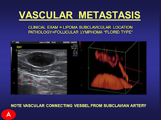
• Subcutaneous “fatty” tumor detected as lymphoma prior to liposuction
• Post PRP scalp swelling/seroma determined to be thrombosed traumatic AV fistula prior to needle aspiration
• Post facial "thread tightening" hemorrhage rediagnosed as bacterial cellulitis
IMAGING ASSISTS SURGICAL PLANNING OF INDICATED BIOPSIES
In my extensive career as the medical director of an advanced imaging diagnostics practice, I have provided great assistance to many surgeons with my work using advanced Doppler Scanning of Tumors and Cosmetic Disorders. I have uncovered countless dermal and subcutaneous issues that would have otherwise gone undetected with less effective technologies, leading to potential complications in the surgical procedure and patient recovery. The advancement in this innovation empowers any upcoming surgical procedure with remarkable confidence of a safer end result. Where biopsies are becoming a thing of the past, our non-invasive 4D Digital imaging replaces weeks of lab work and radiologic tests and often provides more useful information.
DIGITAL BIOPSY CASES: WHAT ARE YOU ABOUT TO BIOPSY? WHAT HAPPENS AFTER THE NEEDLE INSERTS?
Here we have 2 subdermal masses which are mobile, non tender and firm without history of trauma.
Case A: The oval mass (dark echoes=suspicious) with irregular vessels (red) was referred as a probable cyst or lipoma. The tumor is highly vascular and connected from the aorta by way of the subclavian feeding artery. Liposuction would result in massive hemorrhage and spread of tumor cells into the circulation.
Case B: The ovoid white region ( bright echoes=benign) is ossified as confirmed by the CT scan of the coccyx. The sonogram allows you to reassure the patient it is NOT CANCER. It prompts one to avoid a standard needle that would bend, crack or dislodge into the soft tissues requiring exploration to locate/retrieve the broken metal fragment.
For more information or to discuss the many benefits of Digital Pre-Op Imaging, contact us directly at: 212.355.7017 or email: appt@barddiagnostics.com
You can also find us on: Linkedin
by: Dr. Robert Bard
As health practitioners, we are so fortunate to be part of an era where information and technology is at its highest; where our performance is strongly advanced in accuracy, response and safety. This has been made possible due to artificial intelligence (AI) innovations in digital diagnostic scanning equipment.
Let it be scanning potential cancer tumors & malignant disorders or implementing additional 'detective work' on a curious anomaly on or under the skin, the use of 4D Doppler Sonographic technology captures so much more information with absolute precision and accuracy than the latest MRI's, X-rays and CT Scans, and we cover more ground in REAL-TIME (give or take 5 minutes).
For all my friends in the practice of Cosmetic Surgery, DIGITAL PRE-OP is a highly useful stage for many patients who may carry hidden issues that can turn into a pandora's box of complications. I have performed this vital service for European plastic surgeons since 2001 in their centers while currently performing domestically as a digital diagnostics partner for serious physicians and surgeons fulfilling similar needs.
Pre-operative imaging is widely performed to verify tissue planes and measure fat depth. Since patients may have forgotten prior treatments, new scans sometimes reveal extensive sub-dermal calcium deposition, unsuspected fluid collections or thick fibrosis distorting the expected anatomy. Anatomic variants may be observed and avoided. Moreover, patient confidence is enhanced by the extra care provided by this advanced technology.
Some of the most common POST-PROCEDURE COMPLICATIONS include:
- Suture loosening and granuloma formation following blepharoplasty
- Lipoatrophy or fat necrosis following PRP or abdominoplasty
- Filler complications and implant migration
- Doppler verification of vascular compromise (venous or arterial) following facial therapies allowing immediate intervention to prevent blindness or tissue necrosis
CLINICAL LANDMINES
• Foreign body reaction created pigmented lesion simulating malignant melanoma
• Subcutaneous “fatty” tumor detected as lymphoma prior to liposuction
• Post PRP scalp swelling/seroma determined to be thrombosed traumatic AV fistula prior to needle aspiration
• Post facial "thread tightening" hemorrhage rediagnosed as bacterial cellulitis
BENEFITS OF DIGITAL PRE-OP IMAGING
1)Targeted biopsies means less scar formation: Sonogram differentiates cysts / lipomas / sebaceous hyperplasia from cancer
2) Blood vessel mapping for improved preoperative planning: Aberrant glabellar/periorbital vessels detected prior to filler injection/fat transfer
3) Healthy tissue spared for better cosmetic appearance: 3D/4D real time imaging guides operative intervention
4) Fat depth diagnosis leads to optimized thermal treatments: Unsuspected veins diagnosed / avoided prevents dvt-thrombosis
5) Treatment follow up for early assessment of effect: Postop seroma / inflammation / hemorhage / necrosis diagnosed and scanned serially with non invasive modality
6) Midline subcutaneous lesion investigation: Sinus pericranii/sacral cystic connection to nervous system
7) Foreign body localization avoids surgical exploration: Tissue reaction may produce changes mimicking focal lesion and foreign bodies quickly removed under direct visualization
..................................................................................................................................................2) Blood vessel mapping for improved preoperative planning: Aberrant glabellar/periorbital vessels detected prior to filler injection/fat transfer
3) Healthy tissue spared for better cosmetic appearance: 3D/4D real time imaging guides operative intervention
4) Fat depth diagnosis leads to optimized thermal treatments: Unsuspected veins diagnosed / avoided prevents dvt-thrombosis
5) Treatment follow up for early assessment of effect: Postop seroma / inflammation / hemorhage / necrosis diagnosed and scanned serially with non invasive modality
6) Midline subcutaneous lesion investigation: Sinus pericranii/sacral cystic connection to nervous system
7) Foreign body localization avoids surgical exploration: Tissue reaction may produce changes mimicking focal lesion and foreign bodies quickly removed under direct visualization
IMAGING ASSISTS SURGICAL PLANNING OF INDICATED BIOPSIES
In my extensive career as the medical director of an advanced imaging diagnostics practice, I have provided great assistance to many surgeons with my work using advanced Doppler Scanning of Tumors and Cosmetic Disorders. I have uncovered countless dermal and subcutaneous issues that would have otherwise gone undetected with less effective technologies, leading to potential complications in the surgical procedure and patient recovery. The advancement in this innovation empowers any upcoming surgical procedure with remarkable confidence of a safer end result. Where biopsies are becoming a thing of the past, our non-invasive 4D Digital imaging replaces weeks of lab work and radiologic tests and often provides more useful information.
DIGITAL BIOPSY CASES: WHAT ARE YOU ABOUT TO BIOPSY? WHAT HAPPENS AFTER THE NEEDLE INSERTS?
Here we have 2 subdermal masses which are mobile, non tender and firm without history of trauma.
Case A: The oval mass (dark echoes=suspicious) with irregular vessels (red) was referred as a probable cyst or lipoma. The tumor is highly vascular and connected from the aorta by way of the subclavian feeding artery. Liposuction would result in massive hemorrhage and spread of tumor cells into the circulation.
Case B: The ovoid white region ( bright echoes=benign) is ossified as confirmed by the CT scan of the coccyx. The sonogram allows you to reassure the patient it is NOT CANCER. It prompts one to avoid a standard needle that would bend, crack or dislodge into the soft tissues requiring exploration to locate/retrieve the broken metal fragment.
For more information or to discuss the many benefits of Digital Pre-Op Imaging, contact us directly at: 212.355.7017 or email: appt@barddiagnostics.com
..................................................................................................................................................
Reference:
JOURNAL AMERICAN ACADEMY DERMATOLOGY 2012
IMAGE GUIDED CANCER TREATMENTS Springer Publ. 2014
MELANOMA IMAGING AM
ACAD DERMATOLOGY DENVER 2014
3D/4D DOPPLER SCANS WORLD FEDERATION ULTRASOUND 2015
DERMATOLOGIC CLINICS SYMPOSIUM Elsevier Publ. 2017
MT SINAI DERM/SURG WINTER SYMPOSIUM NEW YORK 2017
AMERICAN INSTITUTE OF ULTRASOUND NEW YORK
2018
Disclaimer & Copyright Notice: The materials provided on this website/web-based article are copyrighted and the intellectual property of the publishers/producers (The NY Cancer Resource Alliance/IntermediaWorx inc. and Bard Diagnostic Research & Educational Programs). It is provided publicly strictly for informational purposes within non-commercial use and not for purposes of resale, distribution, public display or performance. Unless otherwise indicated on this web based page, sharing, re-posting, re-publishing of this work is strictly prohibited without due permission from the publishers. Also, certain content may be licensed from third-parties. The licenses for some of this Content may contain additional terms. When such Content licenses contain additional terms, we will make these terms available to you on those pages (which his incorporated herein by reference).The publishers/producers of this site and its contents such as videos, graphics, text, and other materials published are not intended to be a substitute for professional medical advice, diagnosis, or treatment. For any questions you may have regarding a medical condition, please always seek the advice of your physician or a qualified health provider. Do not postpone or disregard any professional medical advice over something you may have seen or read on this website. If you think you may have a medical emergency, call your doctor or 9-1-1 immediately. This website does not support, endorse or recommend any specific products, tests, physicians, procedures, treatment opinions or other information that may be mentioned on this site. Referencing any content or information seen or published in this website or shared by other visitors of this website is solely at your own risk. The publishers/producers of this Internet web site reserves the right, at its sole discretion, to modify, disable access to, or discontinue, temporarily or permanently, all or any part of this Internet web site or any information contained thereon without liability or notice to you.


